Digital X-Rays
Dental radiographs (X-rays) can provide essential information about oral health. They are an important part of a patient’s dental record. Our office uses computers to help capture, store and transmit dental radiographs. The biggest advantage to digital radiographs is that they use much less radiation than traditional x-rays. Secondly, the quality is much better and we can diagnose dental problems sooner.
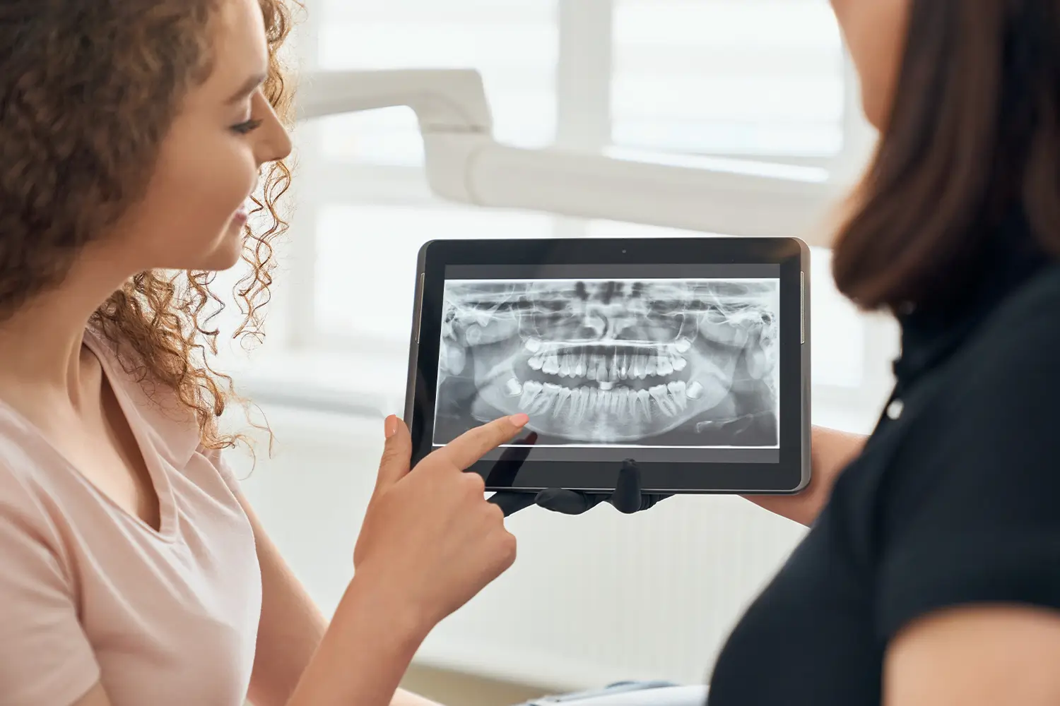
Dental radiographs produced with a special computer create digital images (computerized dental radiographs) that can be displayed and enhanced on the computer monitor.
Creating Digital Radiographs
Digital imaging involves the use of a radiograph machine like that used to create dental radiographs made with film. But instead of using film in a plastic holder, we make digital images using a small electronic sensor or image receptor that is placed in the mouth to capture the image. The x-ray sensor is small computer about the size of a domino that is placed in your mouth in the area to be examined.
When the digital radiograph is exposed, the image is transmitted to a computer processor. Unlike conventional film that may take between three and five minutes to process, a digital radiographic image generally can be viewed quickly on the computer screen. The image is displayed in a large format on the screen, in comparison with the small films that are viewed on a light box. With digital radiographic images, technical errors often can be corrected to provide an optimal radiograph without having to make another exposure. We can use magnification to enhance specific problem areas of a tooth, as well as alter brightness and contrast in the image. Viewing an enhanced dental radiograph on a computer screen can help us better see a problem area.
Digital radiographs eliminate the need for film and film processing chemicals that generate waste, so they are better for the environment.
Frequently Asked Questions
Dental radiographic examinations require exposure to very low levels of radiation (up to 90% less radiation), which makes the risk of potentially harmful effects extremely small.
- small areas of decay between the teeth or below existing restorations (fillings)
- bone destruction from a tooth infection (for example, an abscess) or a cyst;
- bone loss due to periodontal (gum) disease;
- developmental abnormalities;
- some types of tumors;
- the effects of trauma;
- the position of unerupted teeth in children and adults.
- Provides 3-D images of dental structures, soft tissues, nerve paths and bone which are considerably more detailed than conventional two-dimensional dental x-rays
- Allows for more precise diagnosis and treatment planning.
- Is simple and comfortable to take and can diagnostically image both bone and soft tissue simultaneously.
As X-rays pass through your mouth they are mostly absorbed by teeth and bone because these tissues, which are called hard tissues, are denser than cheeks and gums, which are called soft tissues. When X-rays strike the film or a digital sensor, an image called a radiograph is created. Radiographs allow Dr. Melissa to see hidden abnormalities, like tooth decay, infections and signs of gum disease, including changes in the bone and ligaments holding teeth in place.
How often X-rays (radiographs) should be taken depends on your present oral health, your age, your risk for disease, and any signs and symptoms of oral disease you may be experiencing. For example, children may require X-rays more often than adults. This is because their teeth and jaws are still developing. Also their teeth are more likely to be affected by tooth decay than those of adults. Dr. Melissa will review your history, examine your mouth and then decide whether or not you need radiographs.